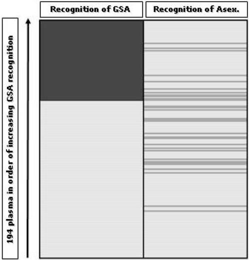
Figure 3. Recognition profiles of plasma IgG from 202 Gambian children.
Erythrocytes harbouring P. falciparum clone 3D7a stage V gametocytes (left column) and asexual parasite stages (right) were tested with each of 194 plasma (rows). Plasma are arranged in increasing order of the proportion of gametocyte recognition events in the right upper quadrant of the flow cytometry dot-blot. Positive antibody recognition is scored as dark grey. Pale fill indicates that antibodies could not be detected above the level of controls (see text).

