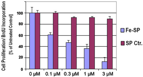
Figure 4. Fe-SP inhibits proliferation of ovarian cancer cells.
Ovarian cancer cells (SKOV-3) were treated with various concentrations (0.1–10 µM) of Fe-SP or non-complexed SP (Ctr.) for 24 h. A colorimetric assay (based on BrdU incorporation detected by a BrdU-antibody peroxidase conjugate) was carried out as described (Materials and Methods). The color intensity at 450 nM correlates directly to the amount of BrdU incorporated into the DNA, which in turn represents proliferation. Experiments were performed in triplicates; data are expressed as the mean of the triplicate determinations (X±SD) in % of absorbance by triplicate samples of untreated cells [ = 100%].

