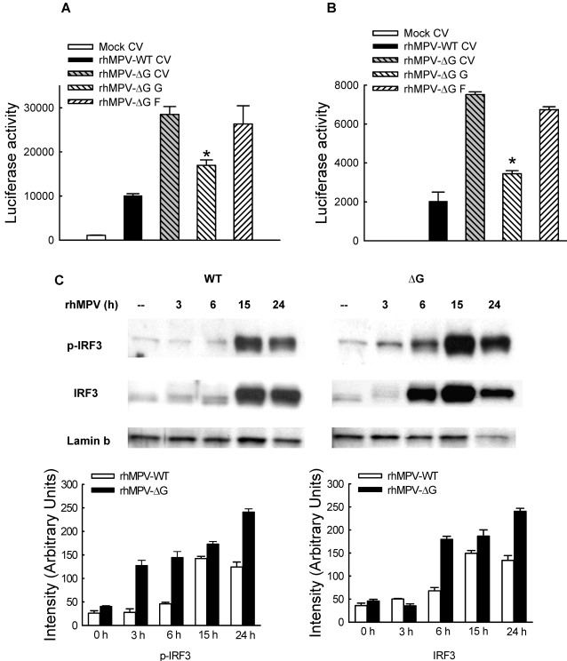
Figure 4. hMPV G protein modulates viral-induced IRF-3 activation.
A549 cells were cotransfected with a luciferase reporter plasmid containing either the human IFN-β promoter (A) or multimers of the RANTES ISRE site (B), and the expression plasmid containing hMPV G or F protein or the control vector (CV), and infected with rhMPV-WT or -ΔG, at MOI of 2. Cells were harvested at 15 h p.i. to measure luciferase activity. Uninfected plates served as controls. For each plate luciferase was normalized to the β-galactosidase reporter activity. Data are representative of two independent experiments and are expressed as mean±standard error of normalized luciferase activity. *, P<0.05, relative to rhMPV-ΔG-infected-CV transfected A549 cells. (C) A549 cells were infected with rhMPV-WT or rhMPV-ΔG, at MOI of 2, for various lengths of time and harvested to prepare nuclear extracts. Equal amounts of protein from uninfected and infected cells were analyzed by Western blot using either an anti-Ser396 phospho-IRF-3 (pIRF-3) or regular anti-IRF-3 antibody. Membranes were stripped and reprobed for lamin b, as control for equal loading of the samples. Densitometric analysis of IRF band intensity, performed using the histogram function of Adobe Photoshop, is shown after normalization to lamin b.

