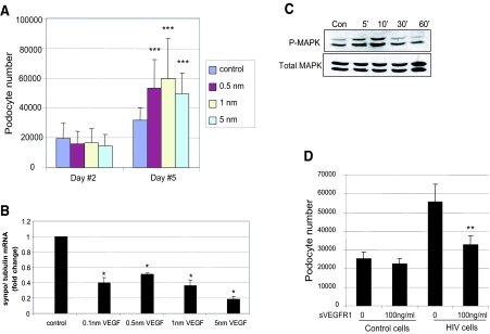
Figure 3.
(A) Effect of VEGF on podocyte proliferation. Podocytes were exposed to no VEGF or VEGF at indicated concentration. Cell number was determined at days 2 and 5. Bars represent mean cell number ± SE of three samples. ***P < 0.005 versus cells without VEGF treatment. (B) VEGF causes reduction of synaptopodin expression. Podocytes were incubated with or without VEGF for 2 d, and RNA was isolated. The synaptopodin/tubulin ratio was determined by real-time PCR. The fold of increase as compared with control podocytes is expressed. Means ± SEM of three independent experiments are shown; *P < 0.05 versus control. (C) VEGF stimulates MAPK phosphorylation. Podocytes were incubated with 1 nm of VEGF or no VEGF for 5, 10, 30, and 60 min. Total and phosphorylated MAPK was determined by Western blot using anti-MAPK and anti–p-MAPK. The representative blot of three independent experiments is shown. (D) Effect of recombinant sVEGFR1 on HIV-induced podocyte proliferation. Control and HIV-infected podocytes were treated with recombinant sVEGFR1 or control vehicle daily for 3 d, and then cell number was determined. **P < 0.01; n = 4 versus vehicle-treated cells.

