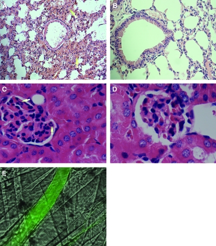
Figure 5.
Anti-MPO IgG induces hemorrhage in specific vascular beds in wild-type mice. (A) Analysis of hematoxylin- and eosin-stained pulmonary tissue sections identified sites of alveolar hemorrhage (arrows) in wild-type mice prestimulated with TNF-α 60 min after anti-MPO IgG infusion (18 μg/g body weight). (B) Well-preserved lung structures after anti-BSA IgG administration. (C and D) No significant structural abnormalities in the glomeruli after the administration of anti-MPO IgG (C; recruited neutrophils indicated by arrows) or anti-BSA IgG (D). Changes in vascular integrity in cremaster muscle microvessels were assessed by measuring the leakage of FITC-conjugated BSA from the microvessels into the surrounding tissue. (E) FITC-conjugated BSA is retained within the venules. Magnifications: ×40 in A and B; ×100 in C and D.

