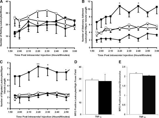
Figure 7.
Effects of Anti-MPO IgG in FcRγ chain−/− mice after intrascrotal TNF-α stimulation. Anti-BSA IgG (○) or anti-MPO IgG (□) was administered intra-arterially (18 μg/g body wt) 2 h after intrascrotal TNF-α (500 ng) injection. The results for mice that received saline intra-arterially 2 h after intrascrotal injection of an elevated concentration of TNF-α (750 ng; ▪) or vehicle (saline containing 0.1% BSA) are also shown (•). (A through C) Changes in leukocyte rolling (A), stationary adhesion (B), and migration (C) were measured for 60 min in postcapillary venules (20 to 50 μm). (D and E) Quantitative MPO immunohistochemistry was performed on pulmonary (D) and renal (E) tissue harvested from TNF-α–pretreated FcRγ chain−/− mice 60 min after the infusion of anti-BSA IgG (□) or anti-MPO IgG (▪; 18 μg/g body wt). Data are means ± SEM (n = 5 for each treatment group). *P < 0.05, **P < 0.01 versus pretreated mice that received intra-arterial anti-BSA IgG (500 ng).

