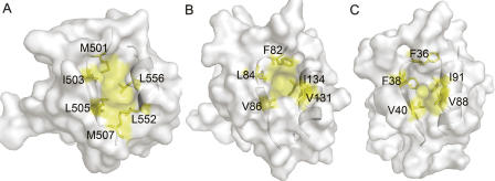
Figure 2.
Comparison of the βB/αB binding pockets. Transparent solvent accessible surface models of PDZ domains, with ribbon representation of βB and αB. Side chains of the hydrophobic residues forming βB/αB binding pockets are labeled and colored yellow; other residues are colored white. (A) Human CASK/LIN-2 PDZ (1KWA). (B) LARG PDZ (2OMJ). (C) GRIP1 PDZ7 domain (1M5Z).

