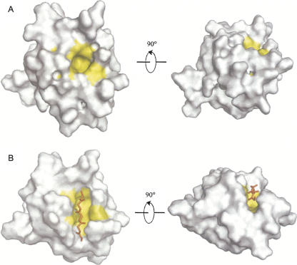
Figure 5.
Surface models of the LARG PDZ in two states highlighting the βB/αB binding pocket. (A) Apo state. (B) Plexin-B1 peptide-bound state. Residues F82, L84, V86, V130, V131, and I134 composing the binding pocket are colored yellow. Shown in the right panels are models rotated (x = −90°) from the left panels for a clear view.

