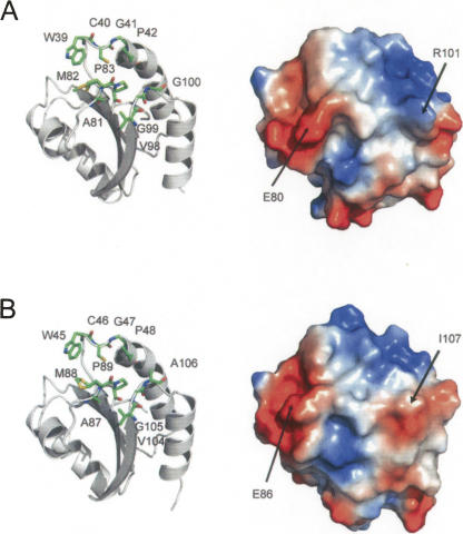
Figure 4.
Substrate-binding loop motif in HvTrxh1 (A) and HvTrxh2 (B). Cartoon display of HvTrxh1 with sticks showing the loop segments Trp39-Pro42, Ala81-Pro83, and Val98-Gly100 (A, left). The vacuum electrostatic potential surface of HvTrxh1 (from the same angle as the left image) with the positions for Glu80 and Arg101 indicated (A, right). HvTrxh2 is presented accordingly (B). Met82 of HvTrxh1 and Met88 of HvTrxh2 are modeled and shown in two alternative conformations. For the HvTrxh2 structure, Cys46 is only shown in the reduced conformation, although it was also modeled in the oxidized conformation.

