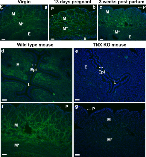
Fig. 3.
Immunostaining of TNX in the uterus (P perimetrium, M myometrium showing longitudinal muscle bundles, M* myometrium showing transverse muscle bundles, E endometrium, Epi epithelium of the lumen, L lumen). TNX (green) is present throughout the uterus of virgin mice (a) and in the uterus during and after pregnancy, shown for uteri at 13 days pregnant (b) and 3 weeks postpartum (c). Cell nuclei are stained with DAPI (blue). TNX is present in the endometrium (d) and the layers of connective tissue ensheathing muscle bundles of the myometrium (f). TNX immunostaining of the perimetrium is relatively weak (a-c, f). The epithelium of the lumen is negative for TNX (d). The specificity of the TNX antibody is demonstrated in e, g. d–g Uteri at 3 weeks postpartum. Bars 50 μm

