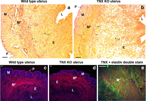
Fig. 6.
Elastin and elastic fibers in the uterus (L lumen). Elastic fibers (purple, modified Hart’s staining) are mostly located in the myometrium (M, M*) and perimetrium (P), whereas the endometrium (E) appears to contain fewer elastic fibers in WT (a) and TNX KO (b) mice. No elastic fiber abnormalities are found in the TNX KO mice. Elastin immunostaining (red) is observed in the myometrium, predominantly in the transverse bundles (M* in c, d). The layers of connective tissue ensheathing the muscle bundles of the myometrium (M) and perimetrium (P) are stained positively for elastin (red). Strong elastin staining is also seen in the endometrium (E). Elastin immunoreactivity is similar for WT (c) and TNX KO (d) mice. Elastin (red) colocalizes (orange) with TNX (green) in the myometrium (M* in e). Not all TNX colocalizes with elastin as TNX also colocalizes with different collagen types (Fig. 4). Bars 50 μm

