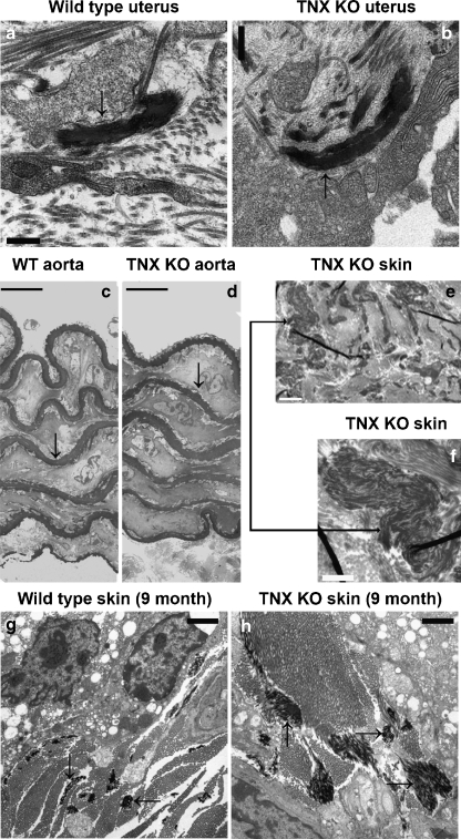
Fig. 7.
Ultrastructural evaluation of elastic fibers. Elastic fibers in WT (a) and TNX KO (b) mouse uterus do not appear to differ in shape or size. The dark structures (arrows) are elastic fibers (shown for uterus at 3 weeks postpartum). The elastic laminae (arrows) in the aorta of WT (c) and TNX KO (d) mice are similar in shape and number (aorta of 9-month-old mice). Skin of older TNX KO mice (9 months old) shows differences in elastic fibers from those of WT skin. Irregular elastin aggregates can be observed in the TNX KO mouse skin (e, near a sebaceous gland). A higher magnification of an elastin aggregate (arrow) is shown in f. These aggregates were not found in skin of 2-month-old TNX KO mice or in 9-month-old WT mice. No irregularities in the shape of elastic fibers are observed in 9-month-old TNX KO mice skin; however, larger elastic fibers than in WT mice (g) are often observed in the TNX KO mice (h, arrows elastic fibers). Bars 0.5 μm (a, b), 10 μm (c, d), 2 μm (e, g, h), 1 μm (f)

