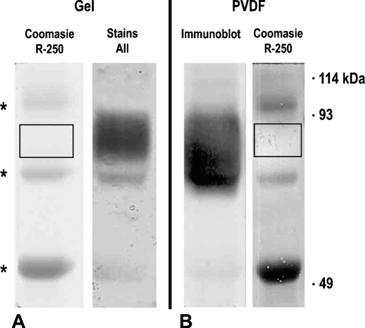
Figure 1.
SDS-PAGE profile of the antigen recognized by the 30B3 antibody. (A) Alternative staining in gel with Coomassie Brilliant Blue or Stains-all. (B) The same material electrotransferred on PVDF membrane and alternatively probed by immunoblotting with the 30B3 antibody (left) or stained by Coomassie (right). Asterisks mark the position of mouse IgG fragments leaking from the column and contaminating the antigen preparation. Note that the 30B3 antigen is not stained by Coomassie. Its position is revealed by Stains-all in gel or immunoblotting on the nitrocellulose membrane.

