Figure 1.
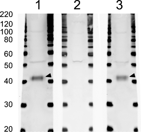
Anti-Ptf1a serum recognizes a 42-kDa protein. Whole cell lysate from embryonic day (e) 15.5 pancreas was loaded in Lanes 1, 2, and 3. In Lane 1, the Ptf1a antiserum recognizes a weak band (∼52 kDa) and a strong band (arrowhead; ∼42 kDa) correlating well with the calculated molecular mass of 37.7 kDa of Ptf1a. In Lane 2, Ptf1a antiserum was preincubated with glutathione-S-transferase (GST)–Ptf1a and the intense 42-kDa band is not detected, whereas the weak 52-kDa band is still present. In Lane 3, the Ptf1a antiserum was preincubated with an unspecific control, GST–Nkx6.1, which does not interfere with the staining pattern as expected.

