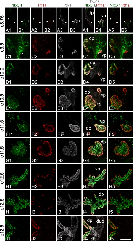
Figure 3.
Ptf1a, Nkx6.1, and Pdx1 localization in the developing pancreas. Whole-mount triple staining was done for Ptf1a, Nkx6.1, and Pdx1 from e8.5 to e12.5 mouse embryos. Stacks of optical sections were recorded on a confocal microscope. Pictures shown in rows A,B,F,G,H,I are single optical sections from a stack, whereas pictures in rows C,D,E,J are composed of several cropped optical sections. This is done to present 3D information in a 2D way avoiding, at the same time, superimposing cells not in the same optical section. All optical sections can be viewed on supplementary movie SM1A–J. Asterisk in F1 indicates that the original Nkx6.1 signal has been bleached by the laser, whereas recording the dorsal pancreas seen in row G thus represents an artifact. (A1–5, B1–5) At ∼e8.75, the first weak Ptf1a-positive cells can be observed in the dorsal pancreas (arrowheads in A1–5). In the ventral pancreas (B1–5), immunoreactivity observed in scattered cells is stronger compared with positive cells in the dorsal pancreas. The first cells positive for Ptf1a appear at ∼e8.5 in the ventral pancreas (data not shown). The dorsal pancreas is devoid of Nkx6.1-positive cells (A1) and Ptf1a expression thus precedes that of Nkx6.1 in the dorsal pancreas. (C1–5) At e9.5, robust Ptf1a expression is observed in the dorsal and ventral pancreas. In the ventral pancreas, Ptf1a is expressed at levels comparable to what is observed later in development. Nkx6.1 is readily observed in the dorsal pancreas. In the ventral pancreas only very few and low Nkx6.1-expressing cells are observed (arrowhead) (see also supplemental movie SM1C). Thus, most Ptf1a-positive cells of the ventral pancreas are negative for Nkx6.1, whereas in the dorsal pancreas most Ptf1a-positive cells are also positive for Nkx6.1. In the dorsal pancreas, Ptf1a- and Nkx6.1-positive cells are found in what appears to be a disorganized pattern, whereas Ptf1a expression pattern in the ventral pancreas is much more uniform. (D1–5) At e10.0, cells positive for Ptf1 and Nkx6.1 are distributed in what appears to be a more organized epithelium compared with e9.5. In the dorsal pancreas, intense Ptf1 and Nkx6.1 immunoreactivity is observed at a level that is comparable to what is seen later in development. Ptf1a and Nkx6.1 positive cells are generally distributed in the bud in a random pattern however, clusters of single positive cells are also present. Nearest to the duodenum, cells tend to be more Ptf1a positive and Nkx6.1 negative/low compared with the rest of the bud, and in some bud areas cells that are Nkx6.1 positive and Ptf1a weak/negative are present. In the ventral pancreas, Ptf1a continues to be expressed at high levels, whereas Nkx6.1 is close to undetectable. (E1–5) At e10.5, Ptf1a, Pdx1, and Nkx6.1 are close to uniformly expressed in the ventral bud and to a large degree in the dorsal bud. Nkx6.1 immunoreactivity reappears in the ventral bud although immunoreactivity is weak. Ptf1a is strongly expressed in both buds. In the dorsal pancreas the tendency of having Ptf1a-positive, Nkx6.1-negative cells nearest to the duodenum continues. Clusters of cells negative for Ptf1a form holes in the otherwise uniform optical sections of Ptf1a-expressing dorsal bud cells. These cells are Nkx6.1 positive. Similar clusters are also observed budding off the Ptf1a-positive area. Such clusters are likely forming endocrine cells. In the rim of the bud, Pdx1 immunoreactivity is more intense compared with the other cells (E3). Furthermore, there is a small tendency for the very center of the dorsal bud to be more Nkx6.1 positive than Ptf1a positive. A few Ptf1a-positive cells can be observed in the duodenum outside the bud area (arrowheads in E4). (F1–5, G1–5) At e11.5, in the ventral pancreas Ptf1a, Nkx6.1, and Pdx1 are coexpressed in almost all cells. Pdx1 is expressed at similar intense levels throughout the bud, but Ptf1a and Nkx6.1 start to display opposite expression levels. This results in a cell population in the rim of the bud that expresses Ptf1a at elevated levels and coexpresses Nkx6.1 weakly, and cells in the center that express Ptf1a weakly and coexpress Nkx6.1 at an elevated level. Expression in the dorsal pancreas is like that of the ventral, just more distinct as Ptf1a expression is extinguished in the very center of the bud, but Nkx6.1 continues to be expressed weakly in the rim cells. In the forming stalk of the dorsal pancreas, Ptf1a and Pdx1 are strongly expressed, whereas Nkx6.1 expression is weaker. (H1–5, I1–5) At e12.5, expression levels and localization of Ptf1a and Nkx6.1 are almost reciprocal, whereas Pdx1 remains more uniformly expressed. Ptf1a is mainly localized to the tips of the branching pancreas where it is expressed at high levels. Nkx6.1 is mainly localized to all cells but the tip cells and the tip cells that do express Nkx6.1 do so at low levels. A few scattered cells express Pdx1 at an elevated level. Such cells are likely forming insulin cells. (J1–5) At e12.5, in the stalk regions Ptf1a-positive cells are localized to the rim of the stalks. Nkx6.1-positive cells are localized throughout the stalk with the weak-expressing cells in the rim and strong-expressing cells in the center. Pdx1 is uniformly localized and expressed. dp, dorsal pancreas; vp, ventral pancreas; duo, duodenum. Magnification is the same in all pictures. Bar = 100 μm.

