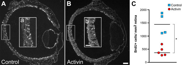
Figure 7.
Decreased BrdU incorporation following activin treatment. BrdU incorporation was assayed in st23 activin treated retinas (A) and control injected retinas (B) using immunohistochemistry. After activin treatment, BrdU incorporation was markedly reduced compared to controls (compare a and b). (C): When quantified, this reduction was found to be significant (* p < 0.05, Mann-Whitney test). Blue squares indicate the means of individual control treated animals and red circles indicate the means of individual activin-treated animals. The medians are indicated by a black line. Scale bars B and b are 100 μm and 50 μm, respectively (valid for A-B and a-b).

