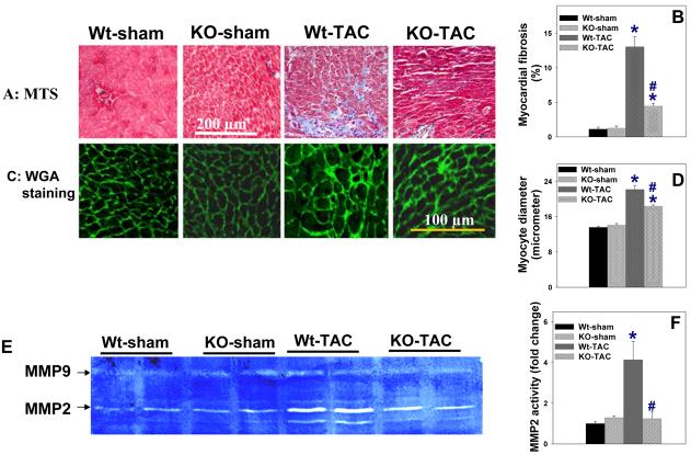
Figure 3.
Histological staining demonstrating that iNOS deletion attenuated TAC-induced myocardial fibrosis (A, B), cardiac myocyte hypertrophy (C, D), and the increase of myocardial MMP2 activity (E,F). MTS: Masson's trichrome staining, blue staining indicates fibrosis; WGA: Staining for wheat germ agglutinin with FITC-conjugated Flur-488 (Invitrogen), bright green staining indicates the area of the matrix and cell membrane. Summarized average data are from 4 representative mice per group. *p<0.05 compared to the corresponding control; #p<0.05 as compared to Wt-TAC.

