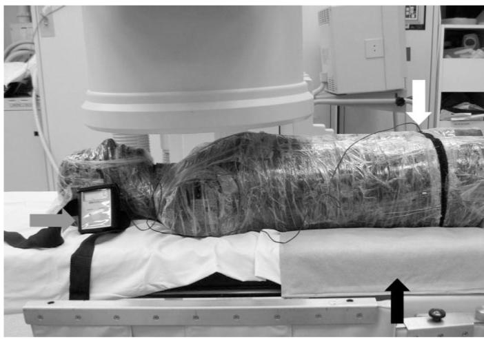
Figure 2.

Scatter radiation to phantom patient ovaries was measured with a dosimeter (gray arrow) by placing the sensor (white arrow) deep in the pelvic cavity with and without a shielding pad. During simulated jugular venous access in the phantom, single and double layers of the shielding pad were placed underneath the pelvic area (black arrow) between the phantom and the table.
