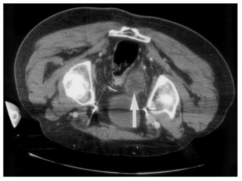
Fig. 3.

Contrast-enhanced CT scan immediately after RFA with the patient in the prone position. The necrotic area in the center of the perirectal tumor enhancement post-RFA, but there is minimal residual enhancement at the periphery (arrow). Pain relief occurred despite residual enhancement.
