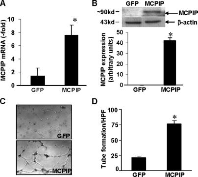
FIGURE 3.
Expression of MCPIP induces capillary-like tube formation in HUVECs. HUVECs were transfected with the MCPIP-GFP expression vector or GFP control for 24 h, and expression of MCPIP was detected by real-time PCR (A) and immunoblot (B) analyses. *, p < 0.001 versus GFP vector-transfected HUVECs. C, phase-contrast photomicrographs (original magnification ×100) of HUVECs seeded on the surface of the polymerized fibrin gels for 24 h after transfection with MCPIP-GFP expression vector or GFP control. D, mean number of tube branch points in randomly selected 5 high power fields (×40) of views was quantified. *, p < 0.05 versus GFP vector-transfected HUVECs.

