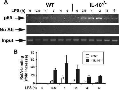
FIGURE 4.
IL-10–/– mice exhibit prolonged LPS-induced RelA (p65) binding to the IL-23p19 promoter after LPS stimulation. A, prolonged LPS-induced RelA (p65) binding in IL-10–/– compared with WT BMDC. The cells were isolated from WT and IL-10–/– mice and stimulated with LPS (5 μg/ml) for the indicated times. ChIP assays were performed using anti-RelA antibodies as described under “Experimental Procedures.” PCR products were separated on a 2% agarose gel and stained with GelStar. The results are representative of three independent experiments. B, Semi-quantitative analysis of RelA binding to the IL-23p19 promoter (–632). WT (white bars) and IL-10–/– (black bars) BMDC were stimulated with LPS (5 μg/ml) at different time points, and ChIP assays were performed as described above. Relative fold changes were determined by semi-quantitative assay using the ABI 7700 sequence detection system as described under “Experimental Procedures.” The results are representative of three independent experiments.

