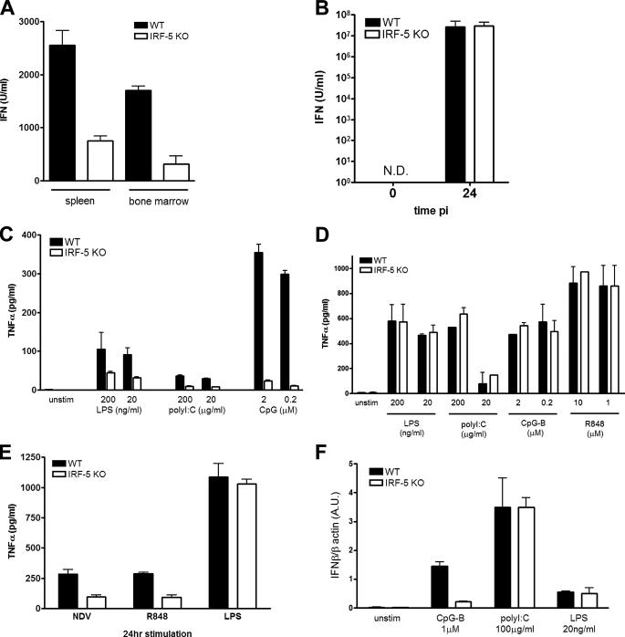
FIGURE 6.
Cell type-specific role of IRF-5 in the antiviral and inflammatory response. A, total white blood cells from spleen and bone marrow cells (2 × 10⁶ cells). B, peritoneal macrophages (2 × 10⁶ cells) from WT and Irf5–/– mice were infected with NDV (50 HAU) for 24 h. The levels of type I IFN in the culture medium were measured by bioassay. C, peritoneal macrophages from WT and Irf5–/– mice were infected with NDV (50 HAU) and treated with R848 (10 μm) or LPS (20 ng/ml) for 24 h, and TNFα levels in the supernatant were measured by ELISA. D, BMDC. E, purified splenic CD11c+ DC (1 × 10⁵ cells) from WT and Irf5–/– mice were stimulated for 16 h with the respective TLR ligands, and levels of TNFα in the medium were measured by ELISA. F, purified splenic CD11c+ DC (1 × 10⁶ cells) from WT and Irf5–/– mice were stimulated for 2 h with respective TLR ligands. Cells were harvested; total RNA was isolated, and relative levels of IFNβ were measured by real time PCR. Transcript levels are shown in arbitrary units (A.U.) compared with β-actin.

