
Fig. 3.
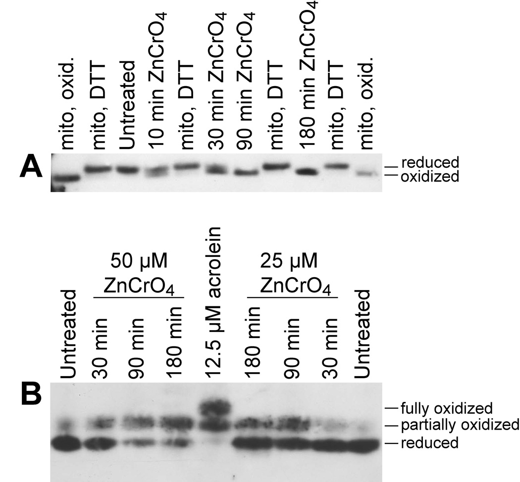
Redox western blots of Trx2 (A) or Trx1 (B) in BEAS-2B cells that were either untreated or treated with ZnCr(VI)O4. In A, cells were treated with 50 µM ZnCr(VI)O4 for the indicated times, and standards for the migration of oxidized Trx2 (“mito, oxid.”) and reduced Trx2 (“mito, DTT”) are included as described in Fig. 1. In B, cells were treated with 25 or 50 µM ZnCr(VI)O4 for the indicated times. Cells treated with 12.5 µM acrolein are included in the center lane to demonstrate migration of the partially and fully oxidized forms of Trx1. The blots shown are representative of replicate experiments.