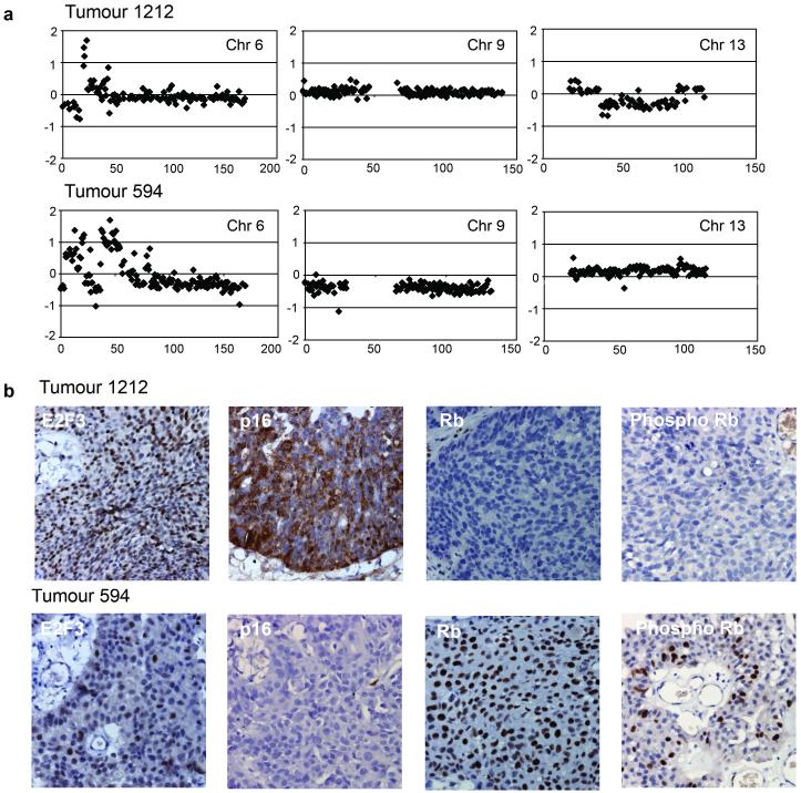
Figure 6.
Array-CGH and immunohistochemical analysis of bladder tumours with 6p22 amplification. (a) Array-CGH analysis of bladder tumours 1212 and 594. Individual chromosome plots of log2 ratio versus distance along chromosome (Mb) are shown for chromosomes 6, 9 and 13. (b) Immunohistochemical analysis of E2F3, p16, Rb and phospho-Rb in bladder tumours 1212 and 594.

