Figure 3.

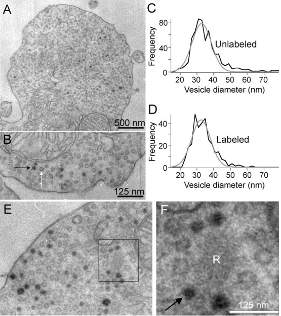
FM dyes taken up during synaptic activity label synaptic vesicles in mouse bipolar terminals. Depolarization-induced uptake of FM1-43 is visible in EM after photoconversion in synaptic vesicles. A, EM image of a synaptic terminal after photoconversion. Some vesicles are filled with dark reaction product. B, Higher-magnification view of the same cell as A. Black and white arrows indicate labeled and unlabeled vesicles, respectively. Sections were not stained with uranyl acetate or lead. C, D, Size distributions of unlabeled and labeled vesicles were identical, with a mean diameter of 33 nm. E, F, Labeled vesicles recycle back to the synaptic ribbon. EM images of a mouse bipolar cell terminal after photoconversion of FM1-43. Black box identifies a synaptic ribbon (R) shown at higher magnification in F. Arrow in F points to one of the four labeled vesicles immediately adjacent to the ribbon. Sections were not stained with uranyl acetate or lead, and so ribbons are pale and filaments that tether vesicles to the ribbon surface are not readily visible.
