
Figure 1.
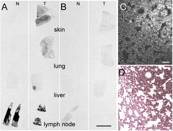
Expression of RACK1 mRNA in pig tissues. (A-B) In situ hybridisation autoradiography of RACK1 antisense (A) and sense (B) probes on depigmented sections. For each probe, normal (N) tissues are displayed on the left (skin, lung, liver and lymph node) and tumoral (T) tissues on the right (cutaneous melanoma, metastatic melanoma from lung, liver and lymph node). Note the intense signal on the tumors compared to the healthy or non-compromised tissues, with the antisense probe, except for lymph node. (C) Darkfield photomicrograph taken from a MeLiM melanoma lung metastasis hybridised with the RACK1 antisense probe. (D) Consecutive section stained with hematoxylin and eosin. The pigmented area in the tumor matches the region which exhibits silver grains on (C). Bar = 1 cm for A and B and 100 μm for C and D.