
Figure 2.
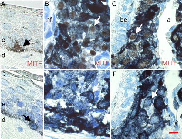
Identification of melanoma cells from MeLiM by MITF. Melanocytes were visualized as brown nuclear granules, by immunohistochemistry with MITF antibody (A-C) and without the primary antibody (D-F). (A, D) Normal skin. (B, E) Cutaneous melanoma. (C, F) Melanoma metastasis in a lung. a, alveolae; be, bronchiolar epithelium; d, dermis; e, epidermis; hf, hair follicle. Arrows point to normal melanocytes (black) and melanoma cells (white). Bar = 100 μm.