
Figure 6.
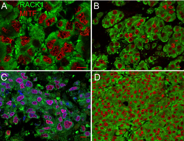
RACK1 in human melanoma metastasis. Confocal microscopy analysis of double labelling of RACK1 protein (green fluorescence) and MITF (red fluorescence). (A, B) Melanoma metastasis in lymph node. (C, D) Melanoma metastasis in liver. High levels of RACK1 are seen in the cytoplasm of all metastatic human melanocytes. Sections are from 4 different patients. Nuclear counterstaining is shown in blue in D. Bar = 10 μm.