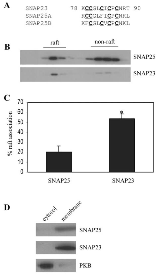
Fig. 2. Comparison of the association of endogenous SNAP-25 and SNAP-23 with lipid rafts in PC12 cells.

A, comparison of the cysteine-rich domains of SNAP-23, SNAP-25A, and SNAP-25B. Cells were solubilized in Triton X-100 and fractionated on a discontinuous sucrose gradient as described under “Experimental Procedures.” Recovered fractions were probed with antibodies specific to SNAP-25 and SNAP-23. B shows representative blots, whereas C is averaged data from five separate experiments. *, p < 0.005. D, distribution of SNAP-25, SNAP-23, and protein kinase B (PKB) between cytosolic and membrane fractions purified from PC12 cells.
