
Fig. 6.

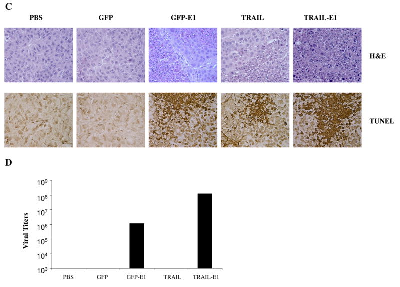
Ad/TRAIL-E1 suppressed tumor growth and prolonged survival in mice bearing radioresistant esophageal adenocarcinoma cells. Seg-1R cells were grown as xenograft tumors in nude mice, and mice bearing tumors that reached 4 to 5 mm in diameter were intratumorally injected with PBS, Ad/CMV-GFP, Ad/TRAIL-F/RGD, Ad/GFP-E1 or Ad/TRAIL-E1 for a total of six injections, with vectors of 5 × 10¹⁰ vp each time. Arrows indicate times when treatments were given. A) Tumor volume was monitored over time after the inoculation of tumor cells. Points, means; bars, SE. ANOVA was performed to determine statistical significant differences between treatment groups. B) Kaplan-Meier survival curves for the treatment groups. C) Representative fields showing hematoxylin and eosin–stained sections (upper) and tumor cell apoptosis by TUNEL assay (lower) in Seg-1R tumors from mice. Mice received the various treatments only once, and tumors were removed 3 days after the treatment. Brown color indicates apoptotic nuclei as visualized using DAB substrate. Apoptotic cells were counted under a light microscope in randomly chosen fields. D) The viral vector replication in supernatants of tumor lysates was determined using the tissue culture infectious dose 50 (TCID50) assay in fresh 293 cells.