
Fig 5. Yki-S168A is hyperactivated.
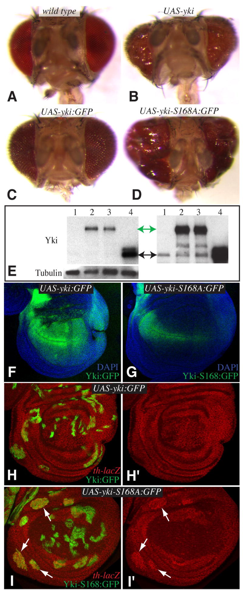
A–D) Heads of adult flies from wild type (A), UAS-yki GMR-Gal4 (B), UAS-yki:GFP GMR-Gal4 (C), UAS-yki-S168A:GFP GMR-Gal4 (D). E) Western blot (4-15% gradient gel) with anti-Yki on lysates from wing imaginal discs of 1. Wild-type 2. sd-Gal4 UAS-yki:GFP 3. sd-Gal4 UAS-yki-S168:GFP. 4. sd-Gal4 UAS-yki. Upper left panel shows a lower exposure, endogenous Yki is barely visible; upper right panel shows a longer exposure of the same gel, endogenous Yki is now visible. Green arrow marks Yki:GFP, black arrow marks Yki. Lower left panel shows anti-α-Tubulin as a loading control. F,G) Wing imaginal discs form sd-Gal4 UAS-yki:GFP (F) and sd-Gal4 UAS-yki-S168:GFP. Images were collected in parallel with same confocal settings, note that Yki-S168A:GFP expression (green) is weaker than Yki:GFP expression but the disc is overgrown. H,I) Wing imaginal discs containing clones of cells expressing UAS-yki:GFP (H) or UAS-yki-S168:GFP (I) (green), and stained for expression of a th-lacZ reporter (red). th expression is induced by yki-S168:GFP (arrows), but not by yki:GFP. In I) th-lacZ expression in the center of the disc is out of the plane of focus.