
Figure 1.
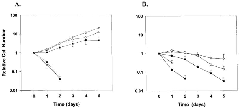
EPO-dependent cell growth in the absence or presence of FCS. Clonal 32D cells lines expressing WT EPOR (open inverted triangle), W282R (closed triangle), YF (open diamond), or 1–321 (closed square), or 32D cells lacking EPOR (closed circle), were maintained in IL-3-containing media. At the start of the assay cells were washed extensively to remove exogenous growth factors and then plated in media containing (A) 0.5 U/mL EPO and FCS or (B) 0.5 U/mL EPO and lacking FCS. Viable cell numbers were determined daily by trypan blue dye exclusion and enumeration. Cell density at individual time points was normalized to the density of the starting population, and the fold increase in cell number was plotted as a function of time in EPO-containing media. Viable cells were not detected in the W282R culture after day 2. Average results from 5 independent experiments are shown.