
Figure 1.
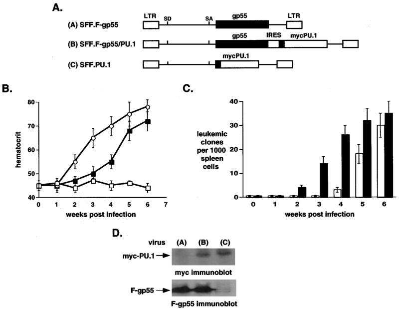
Co-expression of PU.1 and F-gp55 in murine erythroid progenitor cells accelerated both stages of Friend erythroleukemia, in vivo. (a) Schematic representation of retroviral constructs. A myc epitope tag (black box) was placed at the N-terminus of retroviral PU.1. (b) Weekly blood hematocrit levels of retrovirally-infected mice. SFF.F-gp55 virus, filled squares; SFF.F-gp55/PU.1 virus, open circles; SFF.PU.1 virus, open squares. For each virus five mice were infected. Results represent the mean and the standard deviation from the mean. (c) Tumorigenic assays. Mice were infected with SFF.F-gp55 virus (white columns), or SFF.F-gp55/PU.1 virus (black columns). Each week following infection splenic cells were isolated and 1000 cells plated in methylcellulose media without any added cytokines. Ten days following initiation of culture erythroid colonies were identified and scored. Three mice were analysed at each time point. Results represent the mean and the standard deviation from the mean. (d) Immunoblot analysis of splenic extracts from mice infected with viruses. Splenic extracts were prepared from mice 6 weeks following infection with virus: SFF.F-gp55, lane 1, SFF.F-gp55/PU.1, lane 2, and SFF.PU.1, lane 3. Anti-myc immunoblot, upper panel, and anti-F-gp55 immunoblot, lower panel