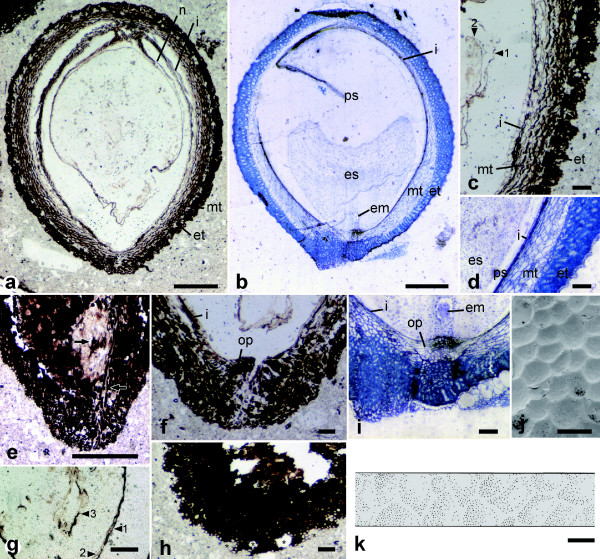
Figure 2.
Fossil and extant seeds of Trimeniaceae share many morphological characters. Section of fossil (a, c, e-h) and seed of Trimenia moorei (b, d, i); surface of T. moorei (j) and fossil (k) seed. Close-up (c) of the micropylar part of (a); (g) is near apex of the nucellus. em, embryo; es, endosperm; et, exotesta; i, inner integument; mt, mesotesta; op, operculum; ps, perisperm. (c, g) arrowheads indicate the nucellar epidermis (1), endosperm membrane (2) and embryo (3). (e) Arrows show tracheids and fibers observed in the hilum. (h) Asterisks indicate areoles seen in nearly paradermal sections of the surface. Scale bars in a and b, 500 μm; e, 200 μm; others, 100 μm.

