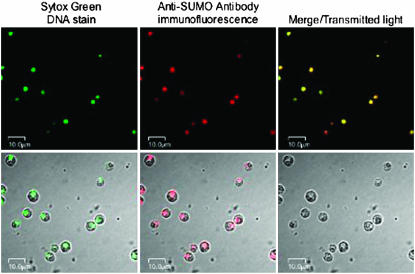
Figure 7.—
In situ localization of CrSUMO96 and its conjugated proteins by immunofluorescence. Wild-type C. reinhardtii cells grown in TAP media were stained with Sytox Green (top left) or detected with anti-CrSUMO96 antibody, which is recognized by the red fluorescent cyanine-5-conjugated goat anti-rabbit antibody (top middle). Images under transmitted light and confocal images are also shown in the top right and the bottom.

