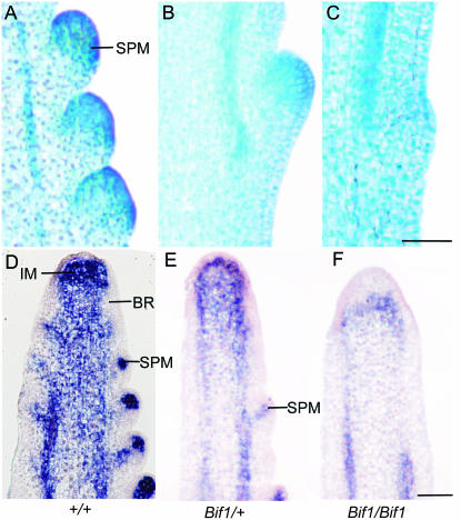
Figure 3.—
Histology and RNA in situ hybridization with kn1 in developing Bif1 tassels. (A–C) Longitudinal sections of 5-week-old tassels stained with TBO, with SPMs visible as areas of intense staining. (A) Three developing SPMs on the flanks of the inflorescence meristem in a normal tassel. (B) Bif1/+, showing a single SPM in the same area as there are three SPMs in normal. (C) Bif1/Bif1 with a slight protrusion on the surface of the rachis but no evidence of developing SPM. (D–F) RNA in situ hybridization with kn1. (D) Meristematic cells and vasculature are indicated by kn1 expression in normal tassels. The absence of kn1 on the flanks of the inflorescence meristem (IM) indicates the formation of the suppressed bract primordia (BR) that subtend SPMs. (E) Bif1/+ inflorescences have fewer areas of kn1 expression on the flanks of the inflorescence. (F) Bif1/Bif1 inflorescence with kn1 expression only in the inflorescence meristem and in the vasculature. Bar, 100 μm.

