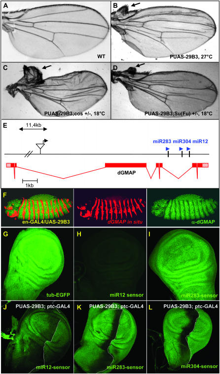
Figure 1.—
The PUAS-29B3 line controls the expression of a microRNA cluster and induces anterior wing outgrowth. (A) Wild-type adult wing and (B) PUAS-29B3/Sd-GAL4 wing obtained at 27°. Note that when flies were grown at 18°, a temperature at which GAL4 activity is much lower, this phenotype is not observed (data not shown). (C) PUAS-29B3/Sd-GAL4; cos2w1/+ and (D) PUAS-29B3/Sd-GAL4; Su(Fu)LP/+ wings obtained at 18°. Arrows indicate outgrowth of anterior wing tissue in the costa region. (E) Localization of the PUAS-29B3 element in gene CG33206, which encodes dGMAP. The microRNA cluster, composed of miR-283, miR-304, and miR-12, is present in the fifth intronic sequence of dGMAP. (F) In situ hybridization for dGMAP mRNA (red), followed by dGMAP immunostaining (green) in PUAS-29B3; en-GAL4 embryos. No increase in dGMAP protein was observed in the en domain. Note that endogenous dGMAP mRNAs are present in all cells but levels are low compared to ectopic RNA expression (middle). Note also that dGMAP proteins are present in all ectodermal cells but their level is artifactually decreased in the en cells where the dGMAP in situ detection is very high (right). (G–L) GFP expression in tub-EGFP (G), miR12-sensor (H and J), miR283-sensor (I and K), miR304-sensor (L), wt discs (G–I), or in PUAS-29B3; ptc-GAL4 discs (J–L). The pictures shown in G, H, and I were obtained with a similar laser setting on the confocal microscope. Note that the laser setting is different for H and J (in which laser intensity has been increased) and thus sensor level is not comparable for these two panels. G–I are illustrative of three independent lines for each sensor. Dotted lines indicate the A/P border. (G–L) Magnification, 200×.

