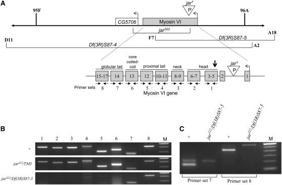
Figure 2.—
Characterization of the genomic region of myosin VI in the Df(3R)S87-5 deletion chromosome. (A) Schematic depicts a portion of the right arm of the third chromosome in Drosophila, showing the position of the myosin VI gene, the deficiencies that remove portions of this region, and the mutations used. The unfilled arrows indicate the transcription start site and direction. Below, an expanded schematic illustrates the exons encoding different parts of the myosin VI protein with eight primer sets (solid arrows) designed to detect different regions (not drawn to scale). The vertical arrowhead indicates the translation start, which is encoded in exon 3. (B) PCR products generated in amplification reactions of genomic DNA using the primers indicated in A, resolved on a 1.8% gel. Size markers (M) are in increments of 100 bp starting with 600 bp at the top. Note that in jar332/Df(3R)S87-5, no PCR products are obtained from exons 3–13, but products are obtained from exons 14–17. (C) RT–PCR products obtained from amplification of total RNA using the primers indicated, resolved on a 1.8% gel. In wild-type animals the higher band is derived from genomic DNA and the lower band from cDNA generated in the RT reaction. Note the lack of the lower band in the mutant genotype. The bands corresponding to genomic DNA amplification were verified by reactions in which the reverse transcription step was not performed (not shown). Size markers (M) are in increments of 100 bp starting with 600 bp at the top.

