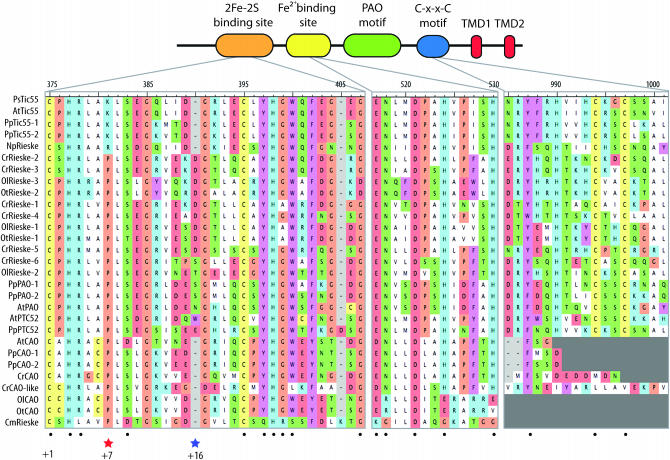
Figure 5.—
Tic55 protein structure. Tic55 overview including the Rieske motif (orange), the mononuclear iron-binding site (yellow), the C-x-x-C motif (blue), two transmembrane domains (red), and the PFAM PAO motif (green). The alignment highlights conserved residues within these domains (black dots). Also indicated are positions where Tic55 orthologs encode a basic residue, instead of a proline at position +7 (red star), and an indel that is absent from Tic55 at position +16 (blue star).

