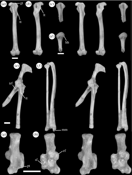
Figure 2.
Stereophotographs of postcranial elements in H. fugax ZMB 50671. Left femora in (a) anterior view and (b) posterior view. Left proximal humerus in (c) anterior view and (d) posterior view. Left pelvis portion and articulated proximal femur in (e) lateral view. Left articulated tibia and fibula in (f) posterior view. Right calcaneum in (g) ventrolateral view and (h) dorsomedial view. Scale bar, 2 mm. Abbreviations: ef, ectal facet; gt, greater trochanter; hh, humeral head; lt, lesser trochanter; mm, medial malleolus; pt, plantar tubercle; sf, sustentacular facet; trf, tuberosity for rectus femoris; tt, third trochanter.

