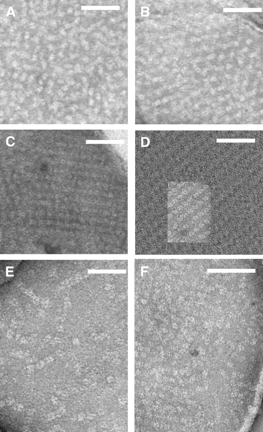
Figure 10.
EM of Negatively Staining Grana Partition Membranes Obtained by Partial Solubilization with α-DM.
(A) to (C) and (E) High-resolution micrographs show the distribution of stain-excluding tetrameric particles: Wild type (A), koCP26 (B), koCP24 (C), and koCP24/26 (E).
(D) A two-dimensional array from koCP24 was superimposed on a larger array from the grana membranes of the barley mutant vir zb63, showing that the crystal lattice is identical in the two samples.
(F) koCP24 periphery membrane areas in which tetrameric particles were less ordered and more widely distributed into a negatively stained background. The bar is 100 nm long.

