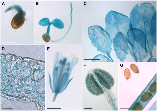
Figure 4.
Analyses of GUS Histochemical Staining in INT1 Promoter/GUS Plants.
(A) and (B) Germinating (A) and young (B) seedling with GUS staining in the cotyledons, the hypocotyl, and the root.
(C) Rosette leaves showing uneven, cloudy GUS staining.
(D) Cross section of a rosette leaf with GUS staining in the mesophyll.
(E) Fully developed flower with staining in all floral organs.
(F) Closed anther with weak GUS staining in all cells and stronger staining in the pollen grains.
(G) Silique with almost mature seeds. No GUS staining is detected in the seeds, but it is detected in all other tissues of the silique.
Bars = 50 μm in (D), 100 μm in (F), 0.5 mm in (A), (B), and (G), 1 mm in (E), and 5 mm in (C).

