
Figure 4. Silencing of uPA expression inhibited tumor growth in an orthotopic mouse prostate tumor model.
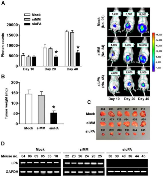
(A) Photon counts of orthotopic prostate tumors on days 10, 20 and 40 (left). Nude mice carrying an orthotopic prostate tumor, established from either mock or siMM-transfected PC3-luc cells, showed substantial bioluminescence signal as indicated by photon counts on day 40. In contrast, the orthotopic prostate tumors developed from PC3-luc cells transfected with siuPA showed significantly low bioluminescence signal (right).(B) Comparison of dissected prostate tumors in (A) from mice 40 days after cell implantation (p<0.01).(C) We excised and photographed prostate tumors in (A) from mice 40 days after cell implantation.(D) RNA samples extracted from PC3-luc prostate tumors (6 animals per group) were analyzed using RT-PCR for uPA expression levels. The siuPA group showed the most prominent and specific knockdown of uPA. GAPDH was used as a control and remained unchanged.