
Figure 6. Histopathology and von Willebrand Factor staining of paraffin-embedded tissue sections from orthotopic prostate tumors.
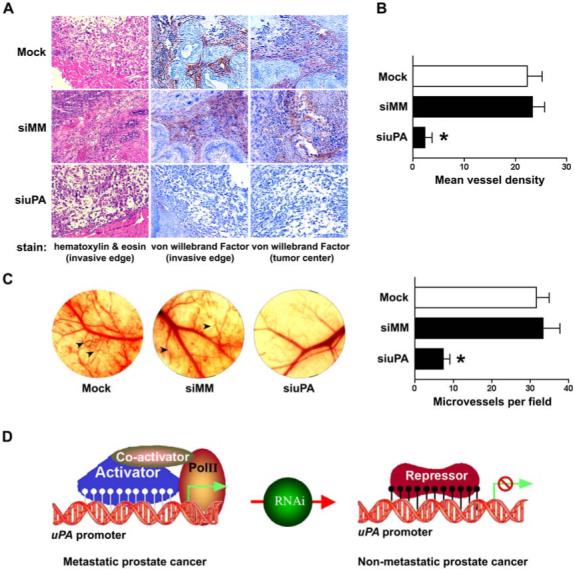
(A) Tumors were harvested from all PC3-luc prostate tumor groups as described in Figure 4(C). Tissue sections (5 μm) were prepared and stained with hematoxylin and eosin (H&E) for histopathological analysis (left column) and with an anti-vWF for immunohistochemistry (middle and right columns). Following blinded procedures, a pathologist examined the H&E and vWF-stained sections (n = 12−15) from each tumor (n = 6 tumors from each treatment group) and determined the morphology, invasive edge and vascularity.(B) Cells positive for vWF counted in five high-power fields in tumor sections with indicated groups. The siuPA group has a significantly lower mean blood vessel density (p<0.01).(C) Tumor angiogenesis was monitored in the dorsal skin-fold chamber of an immunodeficient mouse (left). The photographs show changes in microvessel density of mice skin-folds in response to the secretions of PC3 cells either expressing uPA (mock and siMM) or experiencing uPA knockdown (siuPA). Arrowheads indicate newly formed blood vessels in the skin-folds. Microvessels were counted under a microscope in five random fields (right). The results represent mean ± SD from six implants per group (p<0.01).(D) Schematic model of RNAi-directed alteration of uPA methylation status as a key regulatory switch for metastatic potential.