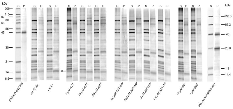
Figure 2.
Total and phosphoprotein staining of immunocaptured complex I. Representative, paired-gel lanes of immunocaptured complex I from HepG2 cells run by PAGE and stained first with Pro-Q® Diamond phosphoprotein gel stain (P lanes) followed by SYPRO® Ruby protein stain (S lanes) as described in materials and methods. HepG2 isolated mitochondria were incubated in phosphorylation buffer (see methods) with the indicated NRTI for 30 minutes before complex I was immunocaptured as described. Pro-Q® stained phosphoproteins of apparent molecular weight of 95, 64, 23.9, and 14.5 kDa were resolved. The 14.5 kDa band (indicated by arrow) showed cAMP/PKAc dependent phosphorylation.

