FIGURE 2.
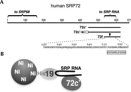
Binding of human SRP72 fragments to SRP RNA. (A) Linear representation of human SRP72. Amino acid residues are numbered. Shown are the N-terminal region involved in the binding to SRP68 and the recently identified RNA binding domain near the C terminus. The SRP72 fragments 72c′, 72c′-ki, and 72f, described by Iakhiaeva et al. (2006) and in this publication, are labeled. The arrow indicates a region that is hypersensitive toward digestion by trypsin. The rectangle indicates a region in 72c′-ki used for radioactive labeling. The boxed sequence shows the Pfam motif (Andersen et al. 2006); X is for any amino acid residue. (B) Schematic representation of the three-component Ni-NTA paramagnetic bead assay showing his-tagged protein SRP19 bound to beads and RNA. Only RNA-bound 72c′ associates with the beads.

