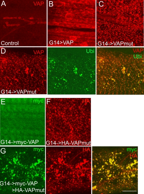
Figure 3. VAPP58S forms ubiquitinated aggregates and induces aggregation of VAPwt in vivo.
(A–C) Confocal images of 3rd instar larval muscles stained with anti-VAP. (A) A control animal (G14-GAL4/+). VAP expression is observed at the neuromuscular junction and in underlying muscles 6 and 7. (B) G14-GAL4/UAS-VAPwt. Increased VAP immunoreactivity is observed with expression of VAPwt. (C) G14-GAL4/UAS-VAPP58S. Punctate staining of ALS-associated mutant VAP is observed. (D) Staining with anti-VAP (red) and anti-ubiquitin (Ubi; green) of animals expressing VAPP58S in the muscle. Mutant VAP forms aggregates that appeared as puncta through out the muscle (red). Ubiquitin immunoreactive puncta also are observed (green) that largely colocalized with the VAP aggregates (merged image). (E–G) Staining with anti-myc (green) and anti-HA (red) of animals expressing myc-VAPwt and HA-VAPP58S in the muscle. (E) Similar to untagged VAPwt and VAPP58S transgenes, expression of myc-VAPwt appears diffuse and cytoplasmic. (F) HA-VAPP58S forms aggregates that appear punctate throughout the muscle. (G) Muscle field of a 3rd instar larva expressing both myc-VAPwt and HA-VAPP58S. Expression of myc-VAPwt (green) appears punctate and colocalizes with HA-VAPP58S aggregates (merged image). Scale bar, 20 µm.

