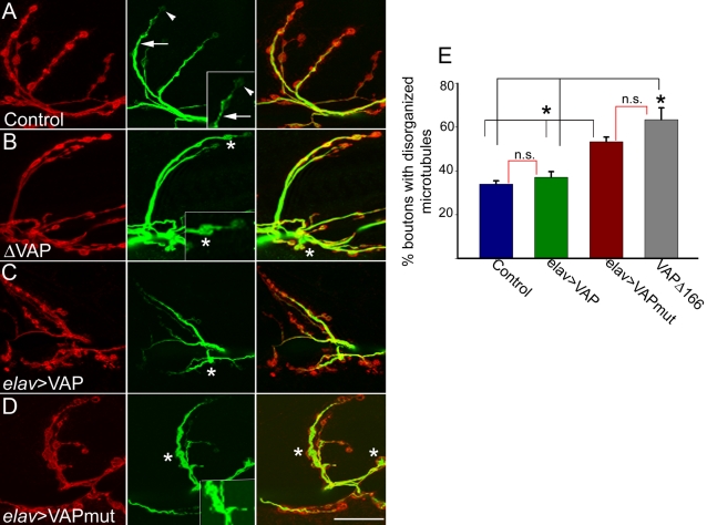
Figure 4. VAPP58S disrupts microtubule organization.
Representative synapses on muscle 6–7 of elav-GAL4/+ (A), VAPΔ166 (B), elav-GAL4/ UAS-VAPwt (C) and elav-GAL4/UAS-VAPP58S animals stained with anti-HRP to recognize the presynaptic membrane (red) and anti-Futsch (green). Anti-Futsch labels stable presynaptic microtubules. (A) In control animals, anti-Futsch staining appears thread-like (arrow). At distal boutons, the staining appears more punctate or diffuse (arrowhead). (B) In VAPΔ166 animals, Futsch expression fills up the cytoplasmic region inside boutons (asterisks). (C) In elav-GAL4/UAS-VAPwt animals, the anti-Futsch staining appears similar to control animals, although some boutons show disorganized microtubule morphology (asterisk). (D) In elav-GAL4/UAS-VAPP58S, more boutons have a disorganized microtubule morphology (asterisk). (E) Quantitation of microtubule morphology assessed using anti-Futsch. The percentage of boutons of the muscle 6-7 synapse exhibiting disorganized microtubule phenotypes was calculated. Values: Controls: 34.0±1.5% (n = 26 synapses). elav-GAL4/ UAS-VAPwt: 37.0±2.6 % (n = 25 synapses). VAPΔ166 (63.4±5.4; n = 15 synapses). elav-GAL4/UAS-VAPP58S (53.3±2.1 %; n = 40 synapses). The black brackets above the histogram indicate all significantly different relationships (*, p<0.001, ANOVA with Kruskal-Wallis test). The red brackets indicate non significant relationships. There was no significant difference in the percentage of boutons with abnormal microtubule morphology between VAPΔ166 and elav-GAL4/UAS-VAPP58S, nor was there a significant difference between elav-GAL4/UAS-VAPwt and the control. Scale bar, 20 µm.

