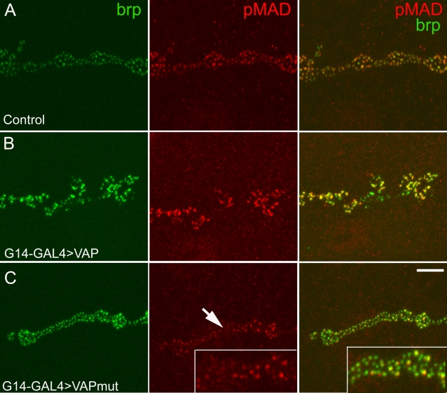
Figure 7. VAPP58S impairs BMP signalling at the synapse.
A–C, representative confocal images of the synapse at muscle 6–7 stained with phosphorylated SMAD (pMAD; red) and bruchpilot (nc82, green), a component of the active zone. (A) In control animals (G14-GAL4/+) postsynaptic expression of pMAD coincides with the expression of presynaptic Bruchpilot as shown in the merged image. (B) In G14-GAL4 /UAS-VAPwt synapses, more intense pMAD staining is observed, indicative of enhanced BMP signalling. (C). In G14-GAL4/UAS-VAPP58S animals, pMAD immunoreactive puncta are less intense (arrow) as compared to the controls indicating reduced BMP signaling. Scale bar, 10 µm.

