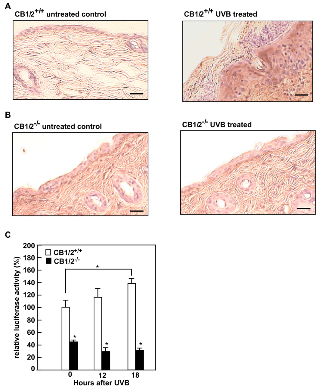
Figure 4.

CB1/2 deficiency suppresses UV-induced NF-κB activation. CB1/2+/+ (A) or CB1/2−/− (B) mice were or were not irradiated with UVB (4 kJ/m2). Dorsal skin from irradiated (right panel of A and B) or non-irradiated (left panel of A and B) mice was harvested 24 h after UVB exposure. Tumor necrosis factor-α (TNF-α) was detected in formalin-fixed and paraffin-embedded sections of skin using the ABC (avidin-biotin complexes) immunohistochemistry staining method. TNF-α is visualized as the brown DAB (3, 3′-diaminobenzidine) staining. All UV irradiation experiments were performed in triplicate for each condition and representative images are shown. The scale bar indicates 25 µm at X200 magnification. (C) CB1/2+/+ or CB1/2−/− MEFs were transiently transfected with 2 µg of NF-κB luciferase reporter plasmid together with 0.1 µg of the pRL-SV-40 plasmid. At 24 h after transfection, cells were starved in 0.1% FBS/DMEM for another 24 h, and then were or were not exposed to UVB (4 kJ/m2) and incubated at 37°C. After 12 or 18 h, the firefly luciferase activity was determined in cell lysates and normalized against Renilla luciferase activity. The relative activity is shown as compared to the activity of CB1/2+/+ MEFs without UVB exposure. Data are represented as means ± S.D. of values from triplicate experiments. Significant differences were evaluated using the Student’s t-test (*, p< 0.05)
